Computerized Axial Tomography
The problem of finding out what is inside you is, in fact, very similar to the problem faced by our milk deliverer in the introduction. Until relatively recently, if you had something wrong internally, you had to have an operation to find out what the problem was. Any operation carries a significant risk, especially in the case of problems with the brain. However, this is no longer the case, as doctors are able to use a variety of scanning techniques to look inside patients in a completely safe way.
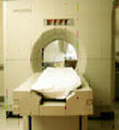
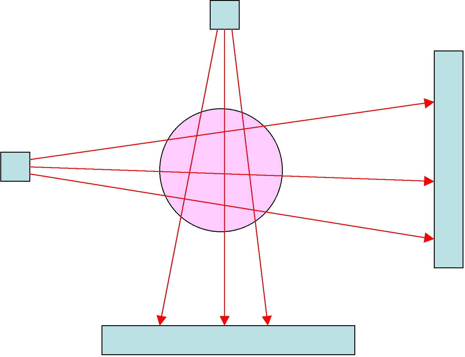
In a CAT scanner, the patient lies on a bed that passes through the hole in the middle of the device. This hole contains an X-Ray source which rotates around the patient. The X-Rays from this source pass through the patient and are detected on the opposite side from the source. The level of intensity of the X-Ray can be measured accurately and the results can then be processed. The resulting fan of X-Rays is illustrated in the following figure (with a conveniently circular patient):
How CAT works
As an X-Ray passes through a patient it is attenuated so that its intensity is reduced. The degree to which this intensity is reduced depends upon what material it passes through. For example, an X-Ray's intensity is reduced more as it passes through bone than as it passes through muscle, an internal organ, or a tumor. A key part in reconstructing an image of the body from a set of X-Ray measurements is making careful measurements of exactly how different materials absorb X-Rays.
When an X-Ray passes through the body, it does so in a straight line. Its total absorption is a combination of the amount that is absorbed by the different materials that it passes through. To see how this happens we need to use a little calculus.